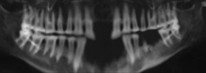
 The VOXEL-MAN program can also calculate simulated X-ray images from the computer tomograms of the mummy’s head. The image shows a panoramic radiograph (or panoramic X-ray) of the teeth, similar to that commonly made by a dentist. It shows some interesting findings:
The radiograph shows not fully dentified upper and lower jaws. In the upper jaw, the two central incisors 11 and 21 are missing. In the lower jaw, the four frontal teeth 32, 31, 41 and 42 are missing. These are no pathological changes, but artefacts due to the embalming of the mummy. In the left lower jaw (in the figure on the right), however, a pathological tooth loss of the teeth 35 and 36 can be seen. From the pre-molar, a part of the root appears at the level of the alveolar crest. At the tip of the root there is a small periapical lightening, which indicates an inflammatory process.
There is a generalized horizontal bone breakdown with vertical bursts, as we know from periodontal changes with bone pockets on the teeth. Abrasions occur throughout the crown area, which can be seen in a clear flattening of the teeth.
Findings: Dr. Andreas Fuhrmann, Polyclinic for X-Ray Diagnostics in Dentistry, Oral and Maxillofacial Surgery, University Hospital Eppendorf.
Note: The denomination of the teeth used here follows the WHO tooth scheme commonly used in dentistry, whereby upper and lower jaws are divided into four quadrants and numbered clockwise. The patient’s right maxilla is designated by 1, the left maxilla by 2, the left mandible by 3 and the right mandible by 4. The second digit indicates the number of the tooth in its respective quadrant, starting from the middle going outwards. For example, the upper central incisor on the right gets the number 11.
Back to the Virtual Mummy