Based on the Visible Human Male, we created a virtual body model of the human torso and internal organs of unprecedented detail and realism. The anatomical and radiological images rendered by the VOXEL-MAN 3D visualization system give an impression of the quality and functionality of the model.
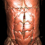 In this view, skin and fat tissue have been removed to show the muscles of the torso, with the rectus abdominis muscle (“six pack”) at center. The images of this series were computed with an illumination by four light sources and shadow casting.
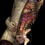 The three-dimensional model of the body consists of more than 650 anatomical structures. The larger organs (bones, muscles, heart, lungs, liver, kidneys, etc.) are represented as voxel objects, the smaller ones like blood vessels and nerves as polygon models. In this image, a midsagittal section reveals some parts of the human anatomy.
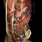 The torso anatomy can be dissected in any direction with any number of cuts. This image shows a layer by layer virtual dissection of different tissue types.
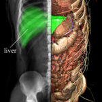 Different viewing modes such as anatomy and X-ray imaging can be combined as desired. The X-ray image can be interrogated at any point to determine which anatomical structures contribute to its intensity.
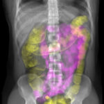 This image shows an X-ray of the complete trunk. Any organ may be highlighted to assess its manifestation in the X-ray image. Here the small intestine is colored in purple, and the large intestine in yellow.
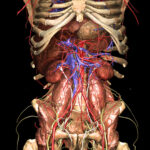 Removal of large portions of the digestive system reveals deeper structures of the body. This view shows the hip bone with psoas and iliacus muscles, as well as various blood vessels such as the abdominal aorta, iliac arteries, and inferior vena cava.
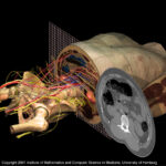 In this view of the internal organs model, a transverse cut reveals the major blood vessels and nerves of the lower abdomen. At any place, the cross-sectional anatomy and its radiological manifestation in computed tomography (CT) can be compared.
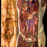 Leonardo da Vinci‘s famous anatomical drawing (around 1500) provided the inspiration for a similar dissection of the Visible Human Male. The image also illustrates the evolution of anatomical knowledge representations from drawings to interactively explorable virtual body models.
Applications
The presented virtual body model provides the basis for the VOXEL-MAN 3D Navigator: Inner Organs atlas of anatomy and radiology, which is available for free download under a Creative Commons license (CC BY-NC-ND). The EUS meets VOXEL-MAN training system for endoscopic ultrasound is another application based on this model.
The 3D model of the Segmented Internal Organs is also available under a Creative Commons license (CC BY).
References
- Karl Heinz Höhne, Bernhard Pflesser, Andreas Pommert, Martin Riemer, Rainer Schubert, Thomas Schiemann, Ulf Tiede, Udo Schumacher: A realistic model of human structure from the Visible Human data. Methods of Information in Medicine 40 (2), 2001, 83-89.
- Andreas Pommert, Karl Heinz Höhne, Bernhard Pflesser, Ernst Richter, Martin Riemer, Thomas Schiemann, Rainer Schubert, Udo Schumacher, Ulf Tiede: Creating a high-resolution spatial/symbolic model of the inner organs based on the Visible Human. Medical Image Analysis 5 (3), 2001, 221-228.
Back to Visible Human Project