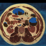
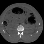
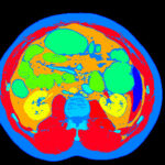
Based on the Visible Human Male published by the U.S. National Library of Medicine, we created a high resolution, multispectral voxel model of the inner organs with more than 200 anatomical objects. We provide this model for research purposes.
Description of the 3D model
The Segmented Inner Organs (SIO) represent a volume of 573×330×774 voxels of 1 mm³ each, covering the torso of the Visible Human Male. The model consists of 774 slices of three TIFF images each:
- cryosection image (24-bit RGB color)
- frozen CT image (8-bit grayscale)
- label image (16-bit numerical object labels)
All images are geometrically aligned. The numbers in the label images indicate to which anatomical object a voxel belongs. The following anatomical objects were segmented and labelled:
abdominal aorta, ampulla, arch of aorta, ascending aorta, ascending colon, azygos vein, body of stomach, bones of the left hand, bones of the right hand, brachiocephalic vein, bronchi, caecum, cardia, cervical vertebra C5, cervical vertebra C6, cervical vertebra C7, coccyx, corpus cavernosum penis, corpus spongiosum penis, cystic duct, descending aorta, descending colon, diaphragm, duodenum (retroperitoneal part), fundus of stomach, gallbladder, greater curvature, grey matter, hepatic veins, inferior mesenteric vein, inferior vena cava, intervertebral disc C6/C7, intervertebral disc C7/T1, intervertebral disc T1/T2, intervertebral disc T2/T3, intervertebral disc T3/T4, intervertebral disc T4/T5, intervertebral disc T5/T6, intervertebral disc T6/T7, intervertebral disc T7/T8, intervertebral disc T8/T9, intervertebral disc T9/T10, intervertebral disc T10/T11, intervertebral disc T11/T12, intervertebral disc T12/L1, intervertebral disc L1/L2, intervertebral disc L2/L3, intervertebral disc L3/L4, intervertebral disc L4/L5, intervertebral disc L5/S1, intervertebral disc S1/S2, ischiocavernosus, larynx, left atrium, left clavicle, left clavicular cartilage, left colic flexure, left costal cartilage 1, left costal cartilage 2, left costal cartilage 3, left costal cartilage 4, left costal cartilage 5, left costal cartilage 6-9, left external oblique, left femur, left hip bone, left humerus, left iliacus, left internal oblique, left jugular vein, left kidney, left lung, left obturator internus, left psoas, left radius, left rectus abdominis, left renal medulla, left renal vein, left rib 1, left rib 2, left rib 3, left rib 4, left rib 5, left rib 6, left rib 7, left rib 8, left rib 9, left rib 10, left rib 11, left rib 12, left scapula, left subclavian vein, left transversus abdominis, left ulna, left ventricle, lesser curvature, liver, lumbar vertebra L1, lumbar vertebra L2, lumbar vertebra L3, lumbar vertebra L4, lumbar vertebra L5, marker 1, marker 2, marker 3, muscles of the left arm, muscles of the right arm, myocardium, pancreas, pelvic diaphragm, penis, pericardium, portal vein, prostate, pulmonary arteries, pulmonary trunk, pulmonary veins, rectum, rectus sheath, right atrium, right clavicle, right clavicular cartilage, right colic flexure, right costal cartilage 1, right costal cartilage 2, right costal cartilage 3, right costal cartilage 4, right costal cartilage 5, right costal cartilage 6-9, right external oblique, right femur, right hip bone, right humerus, right iliacus, right internal oblique, right jugular vein, right kidney, right lung, right obturator internus, right psoas, right radius, right rectus abdominis, right renal medulla, right renal vein, right rib 1, right rib 2, right rib 3, right rib 4, right rib 5, right rib 6, right rib 7, right rib 8, right rib 9, right rib 10, right rib 11, right rib 12, right scapula, right subclavian vein, right transversus abdominis, right ulna, right ventricle, sacrum, scrotum, seminal gland, sigmoid colon, skin of the left arm, skin of the right arm, small intestine, spleen, splenic vein, splenic vein, sternum, stomach, superior mesenteric vein, superior vena cava, testis, thoracic vertebra T1, thoracic vertebra T2, thoracic vertebra T3, thoracic vertebra T4, thoracic vertebra T5, thoracic vertebra T6, thoracic vertebra T7, thoracic vertebra T8, thoracic vertebra T9, thoracic vertebra T10, thoracic vertebra T11, thoracic vertebra T12, thyroid gland, trachea lumen, trachea, transverse colon, unclassified bones, unclassified cartilage, unclassified muscles, unclassified skin, unclassified tissue of the left arm, unclassified tissue of the right arm, unclassified tissue, unclassified veins, urinary bladder, visceral fat, white matter.
License
We are working on a new license model, so distribution of the Segmented Inner Organs is currently paused. Please check back later.
Common questions
The most common questions about the Segmented Inner Organs model are covered in the FAQ.
References
- Karl Heinz Höhne, Bernhard Pflesser, Andreas Pommert, Martin Riemer, Rainer Schubert, Thomas Schiemann, Ulf Tiede, Udo Schumacher: A realistic model of human structure from the Visible Human data. Methods of Information in Medicine 40 (2), 2001, 83-89.
- Andreas Pommert, Karl Heinz Höhne, Bernhard Pflesser, Ernst Richter, Martin Riemer, Thomas Schiemann, Rainer Schubert, Udo Schumacher, Ulf Tiede: Creating a high-resolution spatial/symbolic model of the inner organs based on the Visible Human. Medical Image Analysis 5 (3), 2001, 221-228.
Back to Visible Human Project


